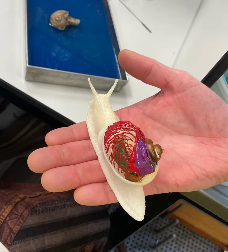
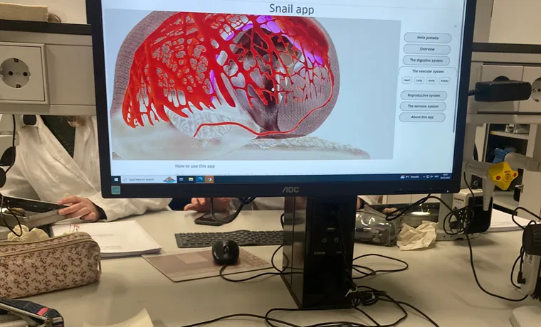
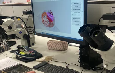
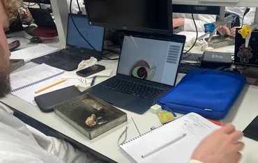
On January 20th, 2025, Anitomical COO Paul Schilperoord was a guest at Wageningen University & Research (WUR) for a dissecting class of the Roman snail (Helix pomatia). In addition to their regular manual and information sources, the dozens of students present were also allowed to consult Anitomical’s prototype application with a digital 3D model and a physical 3D printed model of the Roman snail.
Many students find it difficult to determine the location and orientation of certain organs in the body of the vine snail based on the flat line drawings in the manual. In the real animal, all organs and muscles are now close to or inside each other in the body and have almost the same color. In addition to the manual, there was also a large anatomical model of the Roman snail in the laboratory room, but this is not completely anatomically correct.
Student feedback on the application and the 3D printed model of the Roman snail was largely positive. They indicated that the application with the digital 3D model provides very good insight into what the natural shape of the individual organs looks like, how they are oriented in the body of the snail and where they are located in relation to the other organs.
The reactions to the 3D printed model were also very positive. For some students it is more insightful to handle and view a physical model. The teachers and student assistants who supervised the practical also used the 3D printed model several times to explain answers to questions.
One of the student assistants said in response to Anitomical’s 3D models: “This is really incredibly useful. This makes it so clear. This is really very useful both as an alternative to dissection and as support during the cutting practical. This allows you to see much better what to look for in the snail.”


